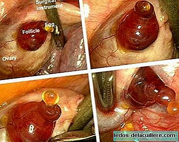
Chance has wanted us to see these amazing images, never captured so far. The photographs show the foreground of an egg emerging from an ovary, and were taken by the Belgian doctor Jacques Donnez, who was performing a hysterectomy when he realized that the patient's ovulation was taking place.
The photographs have been published by the New Scientist magazine, and are the most clear images of a woman's ovulation, so far only the ovulation of animals could be observed.
The images are possible because the mature ovum is a large cell, measures approximately 0.14 millimeters and can be visible to the human eye.
Thanks to these images it has been discovered that, contrary to what was thought, the release of the ovum is not a sudden and explosive event, but that the process takes about 15 minutes.
As the doctor who captured the images points out,
The release of an oocyte (a cell about to become an egg) from an ovary is a crucial event of human reproduction. And these images allow us to have a better understanding of this mechanism.
The process that can be seen in the sequence It is as follows. Human eggs are produced by follicles, sacs loaded with fluids on the surface of the ovary. Shortly after the egg is released, the enzymes separate the tissue from the mature follicle.
This causes the formation of a pink bump and soon after a hole appears from which the egg emerges surrounded by a jelly-like substance that contains cells. And then the egg enters one of the fallopian tubes, which leads to the uterus.
That is, an event that takes place very rarely in the life of a woman, and at very specific moments, so I really consider that these images constitute a milestone in the history of gynecology that will become part of the specialized books, although it really does not involve immediate medical implications.